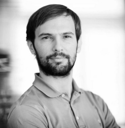
Matteo RauziInstitut de biologie Valrose (iBV) - CNRS / Inserm / Université Côte d'Azur
Mes recherches
My lab is focused in better understanding the mechanisms and unveiling the fundamental principles that govern morphogenesis during the development of the embryo. With a background in engineering and a Ph.D. in developmental biology at the University of Marseille (France, 2010), I joined the EMBL in Heidelberg as a postdoc scientist. My scientific work contributes to a better understanding of the force generating molecular mechanisms driving cell shape changes and the emerging mechanical processes coordinating morphogenesis at the embryo scale. My research combines genetic and optogenetics tools, advanced microscopy, mechanical manipulation, image processing, quantitative analysis and mathematical modelling. In 2017 I was awarded with the ATIP grant and in the same year I joined the CNRS as group leader at University Côte d’Azur in Nice (France) to establish the new team of research “Morphogenesis and mechanics of epithelial tissues”. My lab gathers people from different backgrounds (biology, informatics, physics, and engineering) to generate an interdisciplinary and synergistic team in an international environment.
Mon projet ATIP-Avenir
Subcellular properties and emerging embryo-scale mechanics driving morphogenesis
SPEEMM
The goal of the project is to understand how biophysical forces are generated and how they work to produce exquisitely precise and controlled tissue shape changes in embryo development. Tissue morphogenesis is a process by which the embryo is reshaped into the final form of a developed animal. Tissues are constituted by cells that are interconnected one another: local changes of cell mechanical properties and shape drive consequent tissue shape change. Nevertheless, the knowledge per se of the mechanisms and mechanics at the cell level which drive cell shape changes is insufficient to explain how tissues change their shape. Emerging properties arise at higher scales resulting from the interaction of cells within tissues and of tissues coordinating and interacting with one another. Studying this is a great challenge both technologically and conceptually. I will use the Drosophila melanogaster and the sea urchin Paracentrotus lividus embryo as model systems and focus on the process of tissue folding, process that is vital for the animal since folding defects can impair neurulation in vertebrates and gastrulation in all animals which are organized into the three germ layers. By combining genetic and optogenetics tools, Multi-View light sheet microscopy, mechanical manipulation, image processing, quantitative analysis and mathematical modelling, this project will provide new knowledge on how biophysical forces sculpt functional living organisms.
