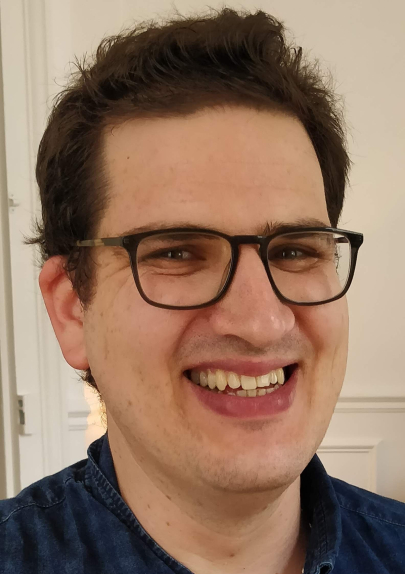
Allon WeinerCentre d'immunologie et des maladies infectieuses (CIMI) - (CNRS / Inserm)
Mes recherches
In my PhD at the Weizmann Institute of Science, Israel (2007−2012) I studied Plasmodium biology using 3D electron microscopy, focusing on nuclear architecture and hemozoin nucleation. In my first post doc in Institut Pasteur (2012−2016), I joined the group of Dr. Jost Enninga to study Shigella invasion using cell biology approaches, functional live imaging and correlative volume tomography. I discovered a new mechanism for vacuolar rupture and outlined the events during early invasion. I also participated in expanding this model to Salmonella invasion. For my second post−doc I joined the group of Dr. Robert Arkowitz in iBV Nice (2016−2018), to study polarized growth of the fungal pathogen Candida albicans. Using dynamic microscopy and 3D EM I revealed that on−site secretory vesicle delivery drives filamentous growth in this organism, in contrast to filamentous fungi and many highly polarised cells. In October 2018 I opened my ATIP-Avenir group in Cimi−Paris, investigating mechanisms of Candida albicans invasion into epithelial layers at the single cell level using a combination of functional live imaging, correlative microscopy and cell biology approaches.
Mon projet ATIP-Avenir
Mechanisms of Candida albicans invasion into epithelial cells revealed by single-cell studies
Candida albicans is an opportunistic fungal pathogen that colonizes the skin, genital and intestinal mucosa of most healthy individuals. In susceptible hosts it can penetrate epithelial barriers and enter the bloodstream leading to severe systemic infection. Infection begins with invasion of epithelial layers, a process thought to occur via an intracellular route. Surprisingly, invasion was also shown to inflict little or no damage to host epithelial cells. These apparently contradictory observations highlight a fundamental gap in our understanding of C. albicans invasion and therefore a need for a detailed account of the mechanisms underlying this process. In our lab we apply innovative single cell functional imaging approaches to uncover the precise path of invasion through epithelial cells and explore molecular mechanisms of invasion, focusing on the role of host cell factors in this process. We also implement advanced 3D correlative light-electron microscopy (CLEM) in the form of “focused ion beam/ scanning electron tomography” (FIB/SEM) to study the precise role of these factors within the complex three-dimensional pathogenhost interface. This project will provide a major step forward in our mechanistic understanding of the enigmatic and medically important process of epithelial invasion.
