Generating symmetry from asymmetric parts
Multicellular epithelia play important roles, especially in the respiratory function. Researchers have identified mechanisms controlling the direction of ciliary beat in such an epithelium. Using an invertebrate model, the planarian, they identified two conserved signaling pathways that cooperate in a distinctive manner to polarize ciliary beat orientation. This work, which resulted in two publications in the journal Developmental Cell, identifies new actors in the polarization of multicellular epithelia. It also sheds light on how animals can generate bilateral symmetry from asymmetric molecules or structures.
Multiciliated cells form up to several hundred motile cilia that beat in a coordinated fashion to generate a flow of liquid or to displace particles and cells. In humans, multiciliated cells are particularly needed for respiratory clearance, a mechanism that renews the protective mucus barrier protecting the lungs from pathogens and dust. Disturbances in the function of these cells caused by certain genetic mutations can thus be at the origin of severe respiratory pathologies.
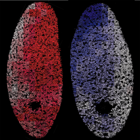
Outside of vertebrates, multiciliated cells are used for the locomotion of diverse animal species such as planarians, flatworms best known for their extraordinary regenerative abilities. These animals also serve as models to understand the function of multicellular epithelia. To fulfill their role, the cilia on the surface of multiciliated epithelia must beat in a coordinated way and in the right direction. In the context of an international collaboration, researchers have shown that in planarians, the ciliary beat is overall directed towards the posterior part of the animal but also towards the lateral edges. This organization generates a symmetrical pattern on either side of the midline, which is obtained via the joint action of two signaling pathways conserved in animals: the Wnt / PCP and Fat / Dachsous pathways.
Unexpectedly, the bilateral symmetry observed in the planarian epidermis is generated from cytoskeletal networks that exhibit chiral asymmetry - an object is said to be chiral if it is not superimposable on its image in a mirror - which derives from the intrinsic chirality of centrioles. The Fat / Dachsous pathway, which controls the mediolateral polarization of the centrioles and the cilia, thus acts via a partially different mechanism in the right and left halves of the body to compensate for the asymmetry of the cytoskeletal network and form a symmetrical pattern.
This work allows to better understand how the so-called Bilaterian animals, to which we belong, can generate a symmetrical organization plan based on chiral constitutive elements, which are widespread in living organisms. It also identifies new conserved molecular actors necessary for the polarization of multicellular epithelia.
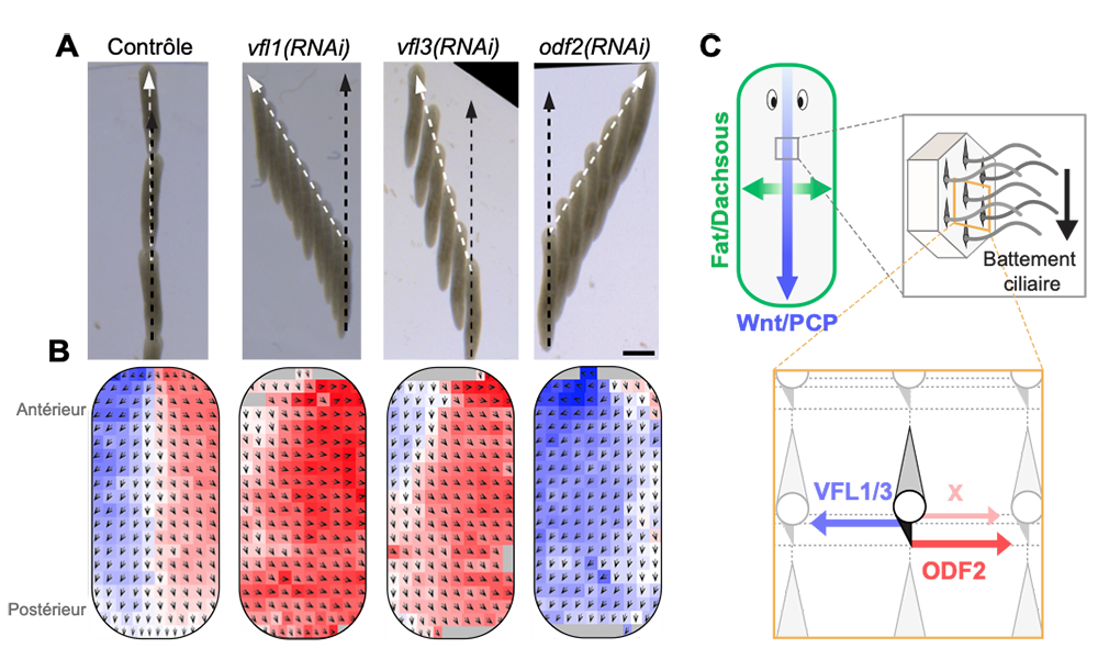
FIGURE. Polarization of cilia in a model of multiciliated epithelium.
A) Effect produced by the inactivation of conserved components of the centrioles - from which cilia are formed and polarized - on the locomotion of planarians. The black dots indicate the theoretical displacement and the white dots the observed displacement. These phenotypes reveal a cryptic chirality of the centriole network mediated by VFL1, VFL3 and ODF2 proteins. B) Analysis of the orientation of the ciliary beat in control or VFL1, VFL3 and ODF2 deficient animals. The black arrows indicate the average values of the direction of the ciliary beat for different regions of the epidermis. These regions are colored red when the beat is oriented to the right and in blue when it is oriented to the left. C) Schemes showing the effect of the Fat / Dachsous and Wnt / PCP pathways on ciliary polarity, and the organization of the cytoskeleton via centriole components VFL1, VFL3 and ODF2. The bilateral symmetry observed in the control animals is obtained by the action of these pathways on a chiral network of centrioles.